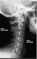
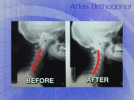
Pre & Post X-ray, MRI and Brain Scan Studies
All images are before and after the successful Atlas Orthogonal Correction, demonstrating how the body can establish a more healthy posture and enhanced nervous system function, achieving a higher state of health.
The areas in the brain scan appearing as holes; before the atlas correction are non-functioning areas of the brain. Function is restored on the post scan.


All images are before and after the successful Atlas Orthogonal Correction, demonstrating how the body can establish a more healthy posture and enhanced nervous system function, achieving a higher state of health.
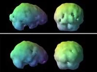
The areas in the brain scan appearing as holes (top view) are before the atlas correction are non-functioning areas of the brain. Function is restored on the one month, post scan. ( bottom view)